Sharing Medical Segmentations in 3D: NIfTI, NRRD, and DICOM-SEG
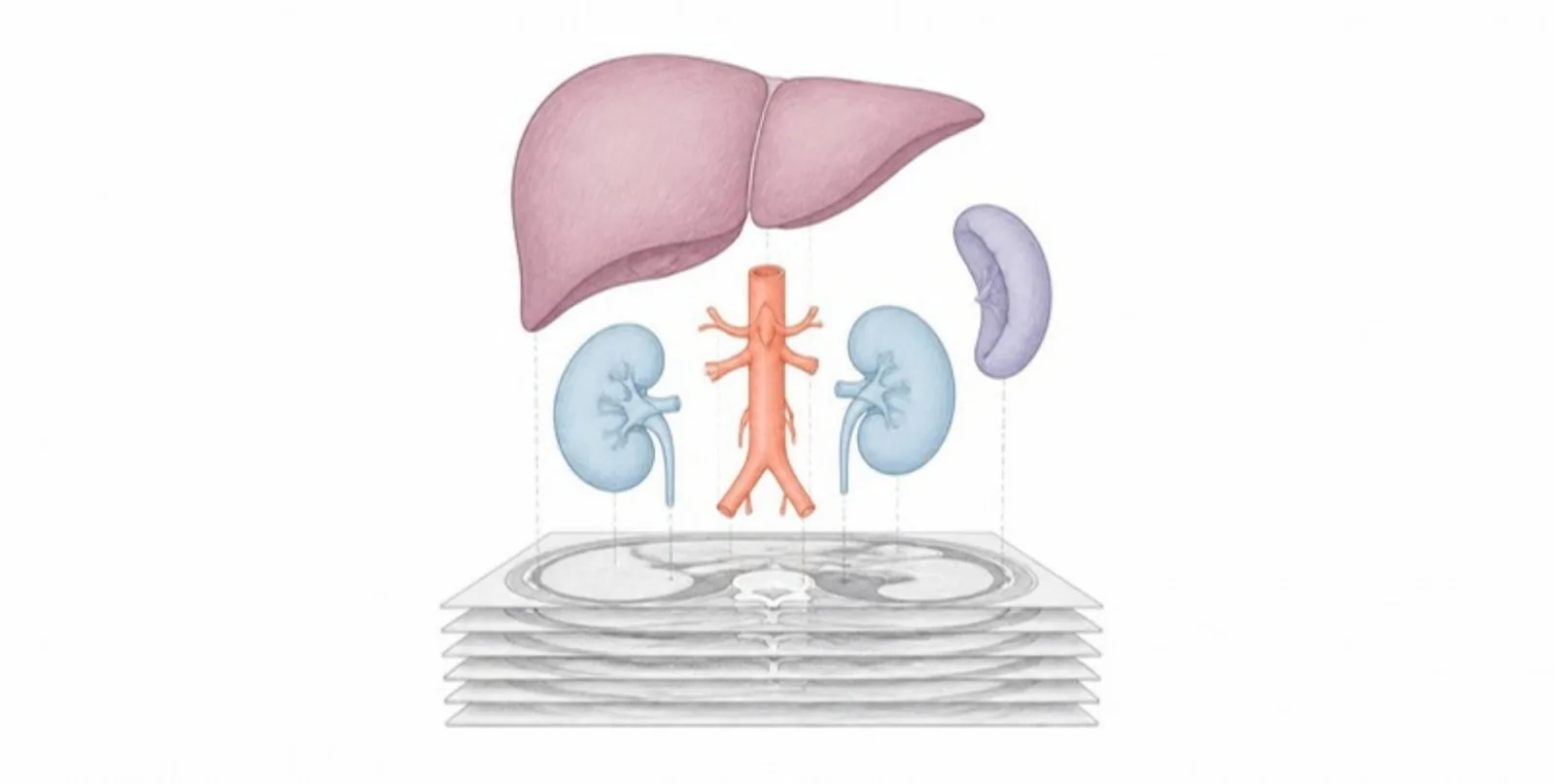
Segmentation is where much of the scientific value in medical imaging lives. Hours of manual contouring or a carefully validated AI model produce label maps that describe anatomy structure by structure — and then they end up as a single screenshot in a figure. Here is how to share the segmentation itself.
Formats you can upload directly
AcademicAR reads the segmentation formats your tools already write:
- NIfTI (
.nii,.nii.gz) — the default for most research pipelines and AI models such as TotalSegmentator or nnU-Net. - NRRD, including 3D Slicer’s
.seg.nrrd— segment names and colours are carried over from Slicer. - DICOM-SEG — the standard segmentation object used by PACS-connected tools.
- A ZIP of masks — one binary mask per structure, as many pipelines export them.
The converter checks the content, not just the extension: a file full of continuous image intensities is recognised as an image, not a segmentation, so you get a clear message instead of a meaningless model.
One structure, one layer
Each non-empty label becomes a separate layer in the viewer, with up to 64 layers per model. That matters for readers:
- They can show, hide, or fade individual structures — hide the ribs to see the lungs, fade the liver to reveal the vessels inside it.
- They can recolour layers or apply a finish, and you can save a default layer view so everyone opens the model the way you intended.
- Names come along where the format provides them, so the legend reads “left kidney”, not “label 7”.
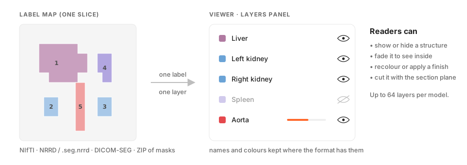 Each non-empty label becomes its own layer that readers can show, hide, fade or recolour.
Large multi-structure outputs are handled one mask at a time, so a full-body TotalSegmentator result does not need special preparation.
Why share the segmentation, not just a render
- Reviewers can check it. Over- and under-segmentation is obvious when you can cut through the model with the section plane.
- AI papers become tangible. Readers see what the model actually segments, in 3D, on their own phone.
- Teaching gets a new resource. A single labelled segmentation becomes an explorable atlas for a whole course.
Privacy still applies
A segmentation can carry identifying metadata, and a skin surface can be recognisable. Use de-identified data, follow your ethics approval, and avoid face surfaces unless you have consent. The uploaded source file is deleted after conversion; only the mesh is kept.
Measurements come built in
For layered models, AcademicAR computes per-structure dimensions and the distances between neighbouring structures — see our post on layer measurements.
Publish your segmentation as an interactive, layered 3D model.