Interactive 3D in Medical Education and Case Reports
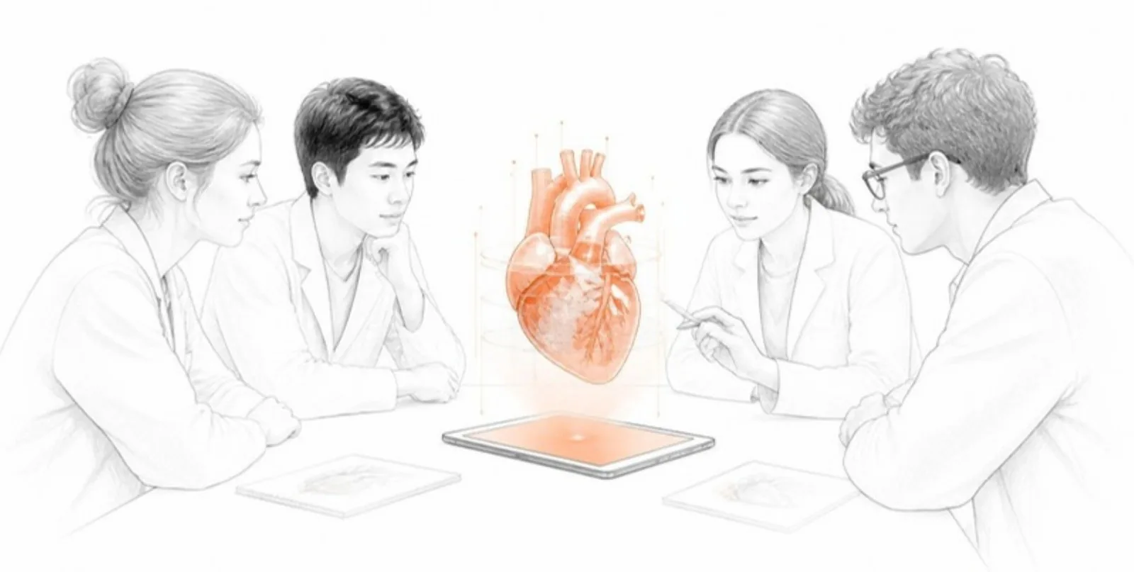
Medicine is taught and published in two dimensions — slides, atlases, journal figures — yet the problems it deals with are spatial. Interactive 3D models built from real imaging close that gap for students, residents, and readers of the literature.
In the classroom and the skills lab
- From slices to anatomy. Students struggle to connect axial CT slices to 3D anatomy. A model with a moving section plane and a colour-coded slice view lets them see both at once.
- Layered dissection. With one layer per structure, students peel away skin, muscle, and bone in order — and rebuild them — as often as they like.
- AR at the bedside of the textbook. On a phone or tablet, the model can be placed on the desk and walked around, with no app to install.
- Guided tours. Save up to 12 views — each with its own camera, visible layers, colours, and section cut — and play them as a step-by-step tour. Each view gets its own link and QR code for handouts. (More in our post on scenes and tours.)
In case reports and clinical research papers
Complex anatomy — congenital heart disease, craniofacial deformities, pelvic fractures, vascular anomalies — is notoriously hard to convey in a few static figures. Linking an interactive model from the figure caption lets readers:
- rotate the anatomy and view it from the surgical approach,
- toggle structures to see what lies behind them,
- check dimensions and distances that the text refers to.
A QR code next to the figure brings the same model to readers of the printed journal or a conference poster.
Doing it responsibly
- De-identify imaging before upload and follow your ethics approval and journal policy for patient images. Facial surface reconstructions deserve extra care.
- Get consent where your institution requires it for publication of case material.
- Be clear about scope. These are communication and teaching tools built from surface reconstructions, not diagnostic or planning software.
AcademicAR makes the compliance step explicit: every upload confirms anonymization, rights, and ethics responsibility, medical uploads carry an extra confirmation, and the raw scan is deleted once the 3D model is built.
For departments
Institutional plans let a department or faculty fund models for all its members from a shared quota, with a public showcase page for its teaching collection. Learn more about institutional plans.
Create your first teaching model from an anonymized scan or a segmentation.